
In-Office Laboratory
Dr. Klyde’s medical office is equipped with the latest, state-of-the-art examination, testing and measurement equipment. This enables Dr. Klyde and his staff to provide you with the best diagnosis and care available, without having to send you through multiple tests and follow-ups. Following are descriptions of some of the examinations that are routinely performed. All of the exams are non-invasive and painless and provide detailed information about your body that will be discussed with you during your visit.

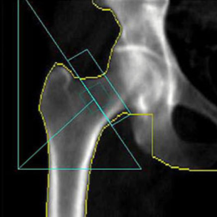
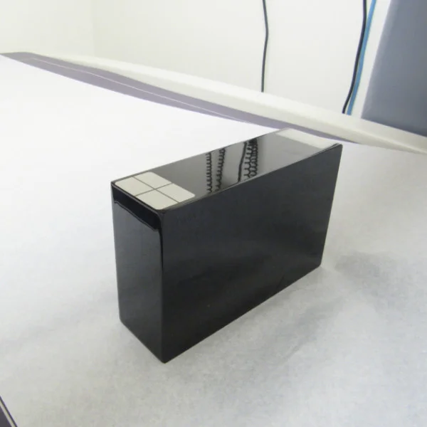
General Electric Lunar DPX Prodigy Bone Densitometer
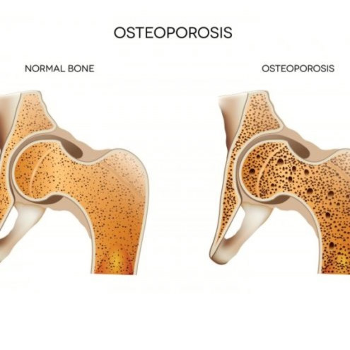
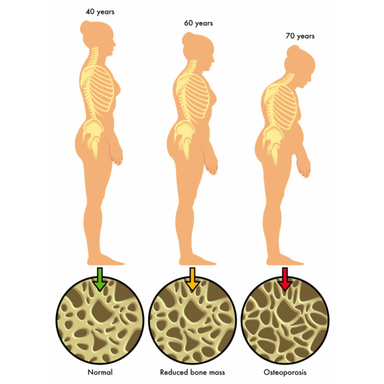
Bone density scanning, also called dual-energy x-ray absorptiometry (DXA) or bone densitometry, is an enhanced form of x-ray technology that is used to measure bone loss. Bone densitometry is an essential tool in osteoporosis management and assessment for metabolic bone disease. It assists physicians in diagnosis, fracture risk assessment, and in monitoring response to therapy. The bone density test itself is painless and quick.
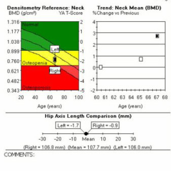
Fracture Risk Assessment
Bone mineral density (BMD) is the strongest tool to predict fracture risk, which increases exponentially as BMD decreases. Femur BMD is recognized as the strongest predictor of femur fracture risk, which has the highest morbidity, mortality and cost of all osteoporotic fractures.

Electrocardiogram (ECG)
The Electrocardiogram (ECG) is the most common test for assessing heart conditions. An ECG is a noninvasive, painless test with quick results. The test consist of electrodes, adhesive attached to the patient’s chest, arms and legs. Each beat of your heart is triggered by an electrical impulse normally generated from special cells in the upper right chamber of your heart (pacemaker cells). An electrocardiogram records the timing and strength of these signals as they travel through your heart. The waveform allows the physician to:
Assess the patient’s heart rhythm
Diagnose poor blood flow to the heart muscle (ischemia, coronary artery disease)
Diagnose a heart attack
Structural problems with the heart's chambers
Diagnose abnormalities of the heart, such as heart chamber enlargement and abnormal electrical conduction
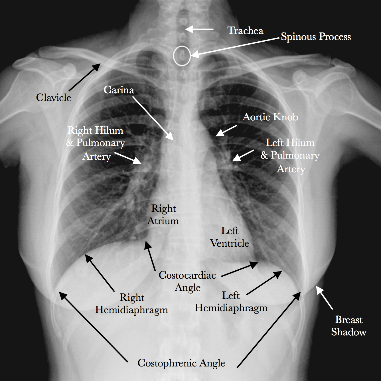

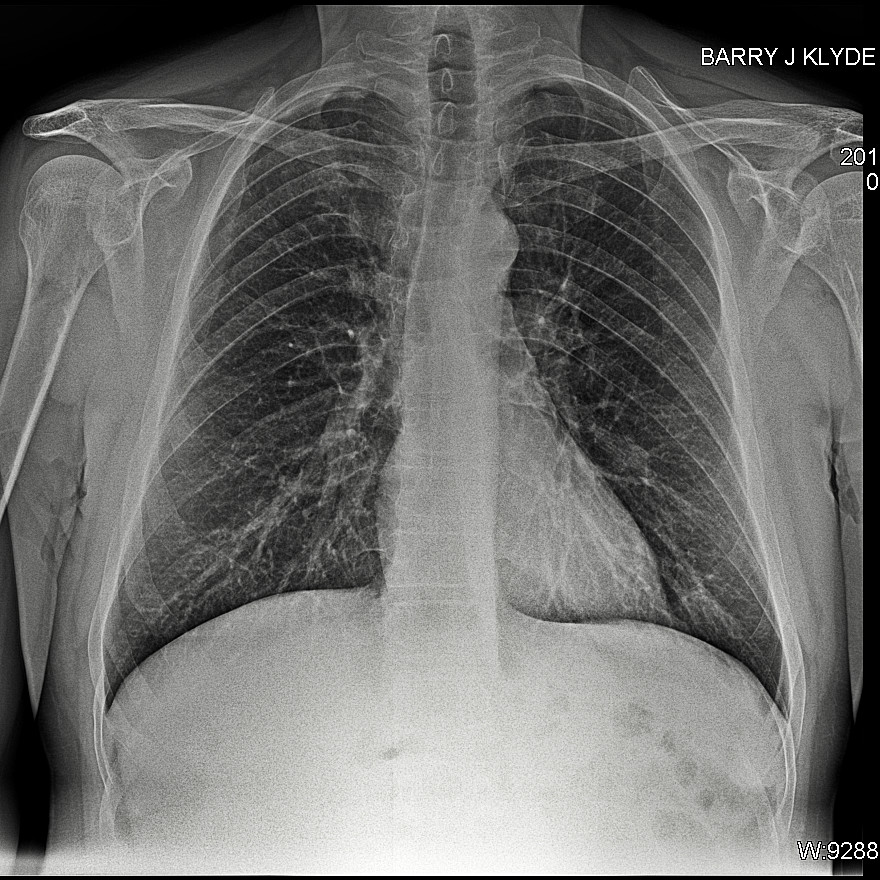
chest x-ray
The condition of your lungs. Chest X-rays can detect cancer, infection or air collecting in the space around a lung (pneumothorax). They can also show chronic lung conditions, such as emphysema or cystic fibrosis, as well as complications related to these conditions.
Heart-related lung problems. Chest X-rays can show changes or problems in your lungs that stem from heart problems. For instance, fluid in your lungs (pulmonary edema) can be a result of congestive heart failure.
The size and outline of your heart. Changes in the size and shape of your heart may indicate heart failure, fluid around the heart (pericardial effusion) or heart valve problems.
Blood vessels. Because the outlines of the large vessels near your heart — the aorta and pulmonary arteries and veins — are visible on X-rays, they may reveal aortic aneurysms, other blood vessel problems or congenital heart disease.
Calcium deposits. Chest X-rays can detect the presence of calcium in your heart or blood vessels. Its presence may indicate damage to your heart valves, coronary arteries, heart muscle or the protective sac that surrounds the heart. Calcium deposits in your lungs are most often from an old, resolved infection.
Fractures. Rib or spine fractures or other problems with bone may be seen on a chest X-ray.
Postoperative changes. Chest X-rays are useful for monitoring your recovery after you've had surgery in your chest, such as on your heart, lungs or esophagus. Your doctor can look at any lines or tubes that were placed during surgery to check for air leaks and areas of fluid or air buildup.
Chest xray heart size, pluminary , lympth nodes and detect masses to determine presesnce of disease. thyroid elagement and trechaea deviation